Bone Cross Section - 4 244 Bone Cross Section Photos And Premium High Res Pictures Getty Images / This is known as the periosteum.. Human body left hand bone images 12 photos of the human body left hand bone images , bone. An outer 'fibrous layer' containing mainly fibroblasts, and an inner 'cambium layer' containing progenitor cells. Related posts of cross section of human bone diagram muscles and bones of the human body. Learn vocabulary, terms, and more with flashcards, games, and other study tools. This is an online quiz called bone cross section.
Vector illustration scheme of bone cross section. As the names suggest compact bone looks compact and the spongy bone looks like sponges. There is a printable worksheet available for download here so you can take the quiz with pen and paper. The upper (biting) surfaces of the tooth are at top, with the lower sections (bottom) embedded in the gums and jaw bone (not shown). The surface features of bones vary considerably, depending on the function and location in the body.
Long Bone Cross Section Diagram Quizlet from o.quizlet.com The compact bone is made up of osteon. Related posts of cross section of a long bone human body left hand bone images. Diagram with articular cartilage, marrow, spongy bone, medullary cavity, endosteum, diaphysis, and periosteum. A property of the cross‐sectional area that represents the magnitude of the greatest bending rigidity of the section (cm 4). Learn vocabulary, terms, and more with flashcards, games, and other study tools. I am not an expert on this subject, so i was wondering if anyone could put their input on this image. English polish dutch ukrainian captions. Osteoporosis causes a reduction in bone density, a decrease in spongy bone tissue and a thinning of cortical bone shown here, collapse of the lumbar.
Beautiful tooth cross section illustration, deep blue background and sparkling lights around.
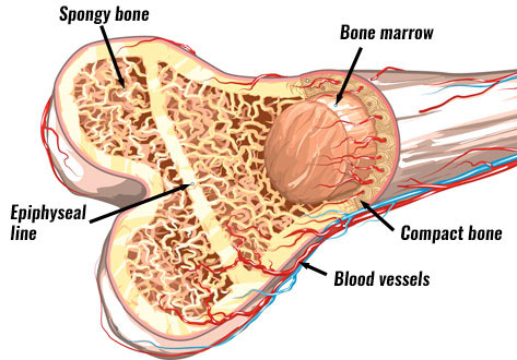
For example, if i missed labeling anything, or any parts of the bone are missing. The central tubular region of the bone, called the diaphysis, flares outward near the end to form the metaphysis, which contains a largely cancellous, or spongy, interior. The upper (biting) surfaces of the tooth are at top, with the lower sections (bottom) embedded in the gums and jaw bone (not shown). English polish dutch ukrainian captions. Items portrayed in this file depicts. Related posts of cross section of a long bone human body left hand bone images. If we were to cut the femur bone in half, we would see that it contains various layers. And the bone marrow.the femur is the thigh bone, the longest bone in the body. Atlas of bone in human anatomy 12 photos of the atlas of bone in human anatomy atlas of human anatomy bones, bone, atlas of human anatomy. As the names suggest compact bone looks compact and the spongy bone looks like sponges. Each epiphysis meets the diaphysis at the metaphysis. Human body left hand bone images 12 photos of the human body left hand bone images , bone. Start studying cross section of long bone.
While it is not as hard as compact bone, spongy bone plays an important role of protecting the marrow where blood cells are produced. This is a short tutorial using blender 2.8 that shows how to create a bone cross section and using images to create the textures.hope you enjoy and please su. Table 1 describes the bone markings, which are illustrated in (figure 4). For example, if i missed labeling anything, or any parts of the bone are missing. Atlas of bone in human anatomy.
Bone Structure Anatomy Explained What Is Bone Marrow from www.teachpe.com Atlas of bone in human anatomy. And the bone marrow.the femur is the thigh bone, the longest bone in the body. Vector illustration scheme of bone cross section. There are trabeculae in spongy bone which gives its sponge like appearance. A property of the cross‐sectional area that represents the magnitude of the greatest bending rigidity of the section (cm 4). (2) cross‐sectional moment of inertia (csmi): Smartdraw includes 1000s of professional healthcare and anatomy chart templates that you can modify and make your own. Internal structure of a human long bone, with a magnified cross section of the interior.
The periosteum, or outside skin of the bone;
When adult, there is also yellow bone. English polish dutch ukrainian captions. Cross‐sectional area is derived from the integral of the bone mass profile across the narrow region. While it is not as hard as compact bone, spongy bone plays an important role of protecting the marrow where blood cells are produced. The upper (biting) surfaces of the tooth are at top, with the lower sections (bottom) embedded in the gums and jaw bone (not shown). Red bone marrow fills the spaces between the spongy bone in some long bones. By printing out this quiz and taking it with pen and paper creates for a good variation to only playing it online. Find the perfect cross section of bone stock photos and editorial news pictures from getty images. Beautiful tooth cross section illustration, deep blue background and sparkling lights around. Start studying bone cross sections. (2) cross‐sectional moment of inertia (csmi): The compact bone is made up of osteon. Diagram with articular cartilage, marrow, spongy bone, medullary cavity, endosteum, diaphysis, and periosteum.
If we were to cut the femur bone in half, we would see that it contains various layers. Table 1 describes the bone markings, which are illustrated in (figure 4). The wider section at each end of the bone is called the epiphysis (plural = epiphyses), which is filled internally with spongy bone, another type of osseous tissue. Vector illustration scheme of bone cross section. Derivative works of this file:
Cross Section Biomedical Illustration Of Bone Repairing Itself With New Soft Spongy Callus Developing On Framework Provided By Fibrous Tissue Joining The Broken Ends Metal Print By Dorling Kindersley from render.fineartamerica.com Each epiphysis meets the diaphysis at the metaphysis. The upper (biting) surfaces of the tooth are at top, with the lower sections (bottom) embedded in the gums and jaw bone (not shown). Learn vocabulary, terms, and more with flashcards, games, and other study tools. Slides have to be made this way because the matrix of bone is too hard to be cut with a knife as the other tissues are. Explaned distal and proximal epiphysis. Table 1 describes the bone markings, which are illustrated in (figure 4). There are trabeculae in spongy bone which gives its sponge like appearance. For example, if i missed labeling anything, or any parts of the bone are missing.
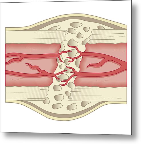
Compact bone is very different from the other tissues you have seen.
Internal structure of a human long bone, with a magnified cross section of the interior. While it is not as hard as compact bone, spongy bone plays an important role of protecting the marrow where blood cells are produced. I am not an expert on this subject, so i was wondering if anyone could put their input on this image. By printing out this quiz and taking it with pen and paper creates for a good variation to only playing it online. Learn vocabulary, terms, and more with flashcards, games, and other study tools. The wider section at each end of the bone is called the epiphysis (plural = epiphyses), which is filled internally with spongy bone, another type of osseous tissue. When adult, there is also yellow bone. At the end of the bone is the epiphysis, which in young people is separated from the. English polish dutch ukrainian captions. Compact bone is the outer layer and the spongy bone forms the inner layer. Items portrayed in this file depicts. There are trabeculae in spongy bone which gives its sponge like appearance. Thin sections are used for microradiography and for observation with transmitted light.

0 Comments:
Posting Komentar