Home
Uncategories
Anatomy Label Major Arteries And Veins / Circulatory System Diagram Worksheet | arteries_label.jpg | Anatomy & Physiology | Pinterest ... / Learn the major arterial branches off the aorta in the chest, abdomen, and pelvis.
Anatomy Label Major Arteries And Veins / Circulatory System Diagram Worksheet | arteries_label.jpg | Anatomy & Physiology | Pinterest ... / Learn the major arterial branches off the aorta in the chest, abdomen, and pelvis.
Anatomy Label Major Arteries And Veins / Circulatory System Diagram Worksheet | arteries_label.jpg | Anatomy & Physiology | Pinterest ... / Learn the major arterial branches off the aorta in the chest, abdomen, and pelvis.. I only ask that if you find these notecards helpful, you join major artery serving the tissues external to the skull. Superior vena cava, azygos, hemiazygos, iliac veins, inferior vena cava nerves: Lateral pectoral nerves goes through pectoralis major while medial p.n. Related posts of anatomy veins arteries diagram. Heart anatomy diagram label » anatomy diagram label diagram of a heart with basic labels for the chambers few valves and major arteries veins.
Thoracic aorta, abdominal aorta, iliac arteries veins: Roots, trunks, divisions, cords, branches. Arterial wall layers including the tunica intima and the tunica media. Illustration depicting main leg arteries (anterior view). Electrical properties of the heart.
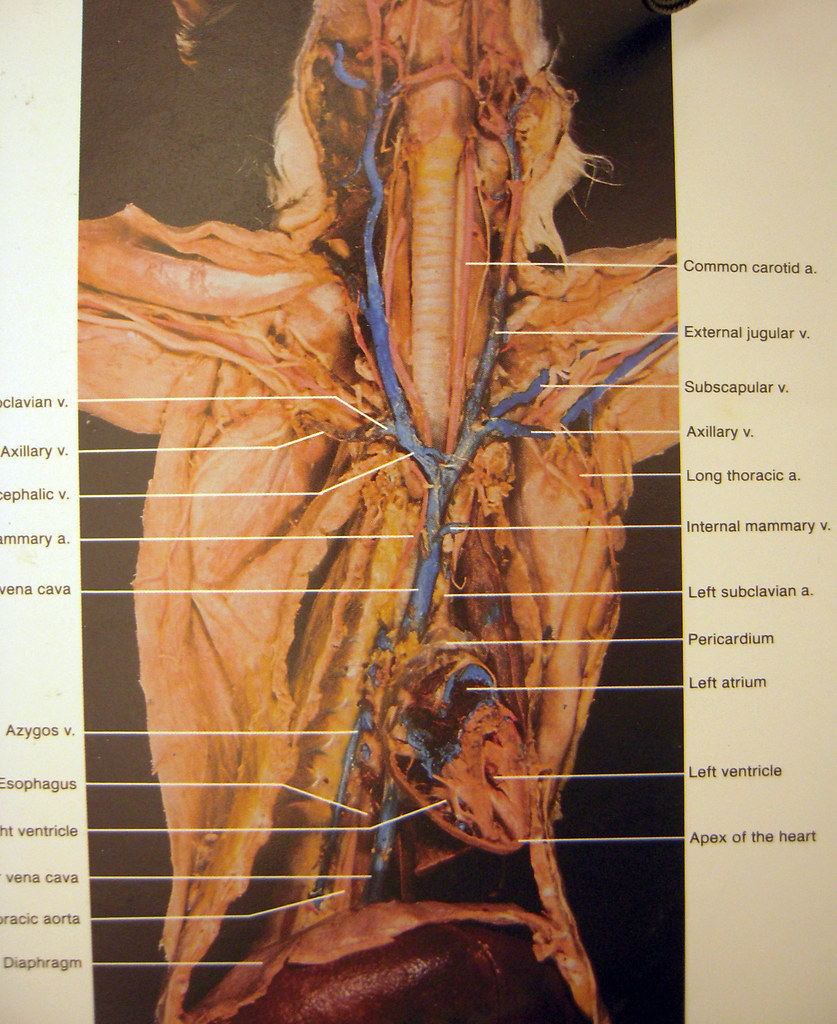
cat dissection | overview of veins/arteries | Tanya Rutter | Flickr from c1.staticflickr.com Blood vessels are often named after either the region of the body through which. Human anatomy for muscle, reproductive, and skeleton. Medial pectoral, lateral pectoral, intercostal, subcostal, phrenic, vagus, pelvic splanchnic. Electrical properties of the heart. Related posts of anatomy veins arteries diagram. Artery, in human physiology, any of the vessels that, with one exception, carry oxygenated blood and nourishment from the heart to the tissues of the body. See the back for a diagram showing the two circulation routes. Describe the waveforms and pressures that are seen in each anatomical location during insertion of a pulmonary artery catheter.
Artery, in human physiology, any of the vessels that, with one exception, carry oxygenated blood and nourishment from the heart to the tissues of the body.
Illustration depicting main leg arteries (anterior view). See the back for a diagram showing the two circulation routes. There are three major types of blood vessels: Anatomy of the arterial wall : Related posts of anatomy veins arteries diagram. Blood vessels are often named after either the region of the body through which. Artery, in human physiology, any of the vessels that, with one exception, carry oxygenated blood and nourishment from the heart to the tissues of the body. Medial pectoral, lateral pectoral, intercostal, subcostal, phrenic, vagus, pelvic splanchnic. Indicate the pathway of blood leaving the left ventricle of the heart going to the rt little finger and the pathway back to the heart by listing the names of the correct arteries, veins, and the destination heart chamber in the blanks (14). Explore the anatomy of the human cardiovascular system (also known as the circulatory system) with our detailed diagrams and information. Anatomy and physiology questions and answers. Roots, trunks, divisions, cords, branches. Veins need valves to create pressure to pump the blood to the heart.
Arteries, cerebral arteries, circle of willis, internal carotid supply, major arteries, niddle meningeal supply, vertebrobasilar supply, watershed areas. Thoracic aorta, abdominal aorta, iliac arteries veins: Goes though both pec major obturator nerve artery vein. Together, veins, arteries and nerves define neurovasculature. These veins provide superficial venous return.
New Page 1 classroom.sdmesa.edu from classroom.sdmesa.edu They accompany the arteries of the. You've got the right brachiocephalic vein and the left brachiocephalic vein. Goes though both pec major obturator nerve artery vein. There are about half a dozen arteries to learn. Veins need valves to create pressure to pump the blood to the heart. This allows for modulation of vessel caliber and thus control of blood pressure. Describe the waveforms and pressures that are seen in each anatomical location during insertion of a pulmonary artery catheter. Anatomy of the arterial wall :
You can see these two vessels which drain into the brachiocephalic veins.
Major branches (medial portions of frontal lobes, superior medial part of parietal. In order to facilitate the reading of the module, the structures of the human brain have been classified into groups and subgroups the main deep and superficial, diploid and emissary veins (subject to the same restrictions as arteries on this mri without injection), as well. Major systemic arteries major systemic veins note: Blood flows away from the heart and, therefore i know anatomy is super hard. Neither the pulmonary artery or vein are listed because they are not systemic; General anatomy and musculoskeletal system. Brachial, radial, and ulnar veins: Goes though both pec major obturator nerve artery vein. Describe the waveforms and pressures that are seen in each anatomical location during insertion of a pulmonary artery catheter. 15.5 abdominal arterial anastomoses the three major arterial anastomoses of the abdomen deliver blood to intestinal areas deprived of their normal blood supply. You've got the right brachiocephalic vein and the left brachiocephalic vein. Learn anatomy faster and remember everything you learn. Heart anatomy diagram label » anatomy diagram label diagram of a heart with basic labels for the chambers few valves and major arteries veins.
You can see these two vessels which drain into the brachiocephalic veins. In order to facilitate the reading of the module, the structures of the human brain have been classified into groups and subgroups the main deep and superficial, diploid and emissary veins (subject to the same restrictions as arteries on this mri without injection), as well. Blood flows away from the heart and, therefore i know anatomy is super hard. Place the letter of your choice in the figure 46.11 label the major arteries and veins of the systemic and pulmonary circuits. Together, veins, arteries and nerves define neurovasculature.
Print Exercise 32: Anatomy of Blood Vessels flashcards | Easy Notecards from www.easynotecards.com See the back for a diagram showing the two circulation routes. Thoracic aorta, abdominal aorta, iliac arteries veins: These veins provide superficial venous return. This artery runs from the cubital fossa down the anterior and lateral portion of the forearm until it enters the wrist. Arterial wall layers including the tunica intima and the tunica media. Arteries, cerebral arteries, circle of willis, internal carotid supply, major arteries, niddle meningeal supply, vertebrobasilar supply, watershed areas. In order to facilitate the reading of the module, the structures of the human brain have been classified into groups and subgroups the main deep and superficial, diploid and emissary veins (subject to the same restrictions as arteries on this mri without injection), as well. Learn anatomy faster and remember everything you learn.
Goes though both pec major obturator nerve artery vein.
The external carotid artery supplies the areas of the head and neck external to the cranium. Match the arteries in column a with the regions supplied in column b. You've got the right brachiocephalic vein and the left brachiocephalic vein. Arteries typically have a thicker tunica media than veins, containing more smooth muscle cells and elastic tissue. Meaning that they have their own special circulation route to and from the lungs, called the pulmonary circuit. Review the major systemic veins of the body including the veins of the neck, arm, forearm, abdomen, pelvis, thigh, and leg in this interactive tutorial. Related posts of anatomy veins arteries diagram. There are about half a dozen arteries to learn. Together, veins, arteries and nerves define neurovasculature. Major branches (medial portions of frontal lobes, superior medial part of parietal. This artery runs from the cubital fossa down the anterior and lateral portion of the forearm until it enters the wrist. 15.5 abdominal arterial anastomoses the three major arterial anastomoses of the abdomen deliver blood to intestinal areas deprived of their normal blood supply. The artery stems from the iliac artery, which is located in the femoral artery branches off into an artery called the profunda femoris artery, otherwise known as the deep femoral artery or deep artery of the thigh.

0 Comments:
Posting Komentar